
Microalgae can provide biomass and valuable compounds for food, biotechnology and other bio-based applications, but producing them efficiently at large scale is more complex than simply supplying light, nutrients and water.
Outdoor cultures respond constantly to changing light and temperature. Too much oxygen can slow photosynthesis. Unfavourable production conditions can stress or damage cells. And unwanted microorganisms can enter the system and compete with the species being produced.
Three recent studies involving researchers from the University of Almería explore complementary ways of addressing these challenges. Together, they show how real-time sensors, advanced microscopy and artificial intelligence can provide a much clearer picture of what is happening inside a microalgae culture and help move production towards more precise and responsive operation.
Why does this matter?
Industrial microalgae production takes place in dynamic biological systems. Conditions can change during the day, cultures can become stressed, and contamination may develop before it is obvious to an operator.
Traditional measurements often provide only part of the picture or require samples to be removed and analysed offline. This creates a delay between a change occurring in the culture and operators being able to react.
Better monitoring could help producers identify problems earlier, adjust operating conditions and use resources more efficiently. The three studies approach this challenge at different levels: one looks at the performance of the reactor, another at the condition of the microalgae, and the third which organisms are actually present in the culture.
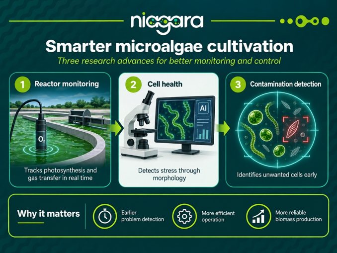
Reading the reactor through oxygen
One study focused on large-scale raceway reactors, one of the most widely used systems for commercial microalgae production because of their relatively simple design and scalability.
During photosynthesis, microalgae take in CO₂ and release oxygen. This makes dissolved oxygen an important indicator of what is happening inside the culture. Too much oxygen can inhibit photosynthesis, while gas exchange also determines how efficiently the reactor can operate.
Researchers developed a method that combines dissolved-oxygen measurements with controlled pulses of air to determine both oxygen production and gas-transfer performance directly during reactor operation. The methodology was tested in an 80 m² outdoor raceway reactor.
The results showed a clear relationship between solar radiation and oxygen production. In practical terms, this means that oxygen evolution can provide information about photosynthetic activity and act as an indicator of biomass productivity. The study also confirmed the importance of the reactor’s sump—the section where aeration is applied—for removing accumulated oxygen.
Rather than periodically taking a sample and waiting for laboratory results, approaches like this could allow operators to follow reactor performance at frequent, regular intervals throughout the day and adjust aeration or other operating conditions in response to what is actually happening in the culture.
Looking at the cells
A second study moves from the reactor scale to the organisms themselves. Arthrospira platensis, commonly known as Spirulina, grows as long filaments. Their length, diameter and structural integrity change depending on the conditions experienced by the culture. This means morphology can provide useful information about whether the organisms are growing normally or experiencing stress.
Researchers used Microdeep™, a microfluidic microscope integrating automated imaging and artificial intelligence, to analyse Arthrospira filaments under different temperatures, pH values, light intensities and agitation conditions.
The system measured filament length and diameter and estimated biomass concentration. Its image-based biomass measurements showed a very strong correlation with conventional gravimetric dry-weight measurements, with an R² above 0.99.
More importantly, the images revealed how the organisms responded when conditions became unsuitable. Under extreme pH, very high light intensity, or temperatures of 10 °C or 45 °C, the filaments showed greater fragmentation, reduced elongation and declining biomass.
This adds another layer of information to conventional process monitoring. A reactor may tell us what temperature, pH or oxygen level it contains; imaging the organisms themselves can help show how the culture is actually responding to those conditions.
Detecting unwanted neighbours
Good production conditions are only part of the challenge. Open and large-scale systems can also be affected by contamination. A third study investigated whether high-resolution holographic reconstruction combined with fluorescence microscopy could identify unwanted Chlorella sorokiniana cells growing alongside Haematococcus pluvialis.
This is important because a fast-growing contaminating species can gradually take over a culture and reduce the production of the desired biomass or compounds matters because a fast-growing contaminant can quietly take over a culture, cutting into production of the desired biomass or compounds. Catching it early gives producers a real chance to intervene before it gets out of hand.
The HOLODETECT HiRes + FLUOR3 system captures information on cell morphology together with fluorescence signals. AI-based models were then trained to recognise and quantify the different species.
The study showed that the system could produce cell counts comparable with conventional microscope counting and quantify the relative abundance of the two species in mixed cultures. However, model design mattered: simpler species-specific models either overestimated total cell counts or struggled to correctly identify which species a cell belonged to, while a model trained to distinguish both species simultaneously provided better discrimination.
The measurements were highly reproducible, with coefficients of variation below 5%. One practical limitation was that very dense cultures of the smaller Chlorella cells needed to be diluted because overlapping cells reduced detection accuracy.
The research therefore does not yet eliminate every practical difficulty associated with online contamination monitoring, but it demonstrates how combining imaging, fluorescence and AI could support earlier and more automated detection of unwanted organisms.
From measurement to smarter production
Taken together, the three studies illustrate different pieces of the same transition. At the reactor level, dissolved oxygen can provide real-time information about photosynthesis and gas exchange. At the organism level, AI-assisted microscopy can reveal morphological signs of growth or stress. At the culture-composition level, holographic and fluorescence imaging can help distinguish the organisms producers want from those they do not.
These technologies do not replace the need for biological expertise or conventional analytical methods. They provide additional information that can potentially be obtained faster and more continuously.
The wider objective is therefore not simply to collect more data. It is to make microalgae production more responsive: detecting changes earlier, understanding what they mean and using that information to make better operational decisions.
For large-scale microalgae production, that could translate into healthier cultures, fewer losses from contamination, more efficient reactor operation and more consistent biomass production.
Ultimately, smarter production starts with being able to see what is happening—inside the reactor, inside the culture and even at the level of individual cells.